Close

Varieties of Neuronal Morphology

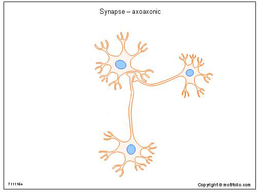
Neuronal Cell Bodies come in many shapes and sizes

Multipolar and Unipolar (or Pseudo-Unipolar) Neurones

Multipolar neurones have many dendrites and have a single axon. the dendrites collect information from different sources and the axon carries action potentials to other neurones or target organs. Motor neurones and interneurones are examples of mulitpolar neurones.

Unipolar (or pseudo-unipolar) neurones have a cell body with a short axon that divides into two after a short distance; these axons travel in opposite directions and function as a single long axon.

The main example of a unipolar neurone is a dorsal root ganglion neurone; one axon has sensory terminals in the skin, muscle, joints or viscera, and the other branch terminates in the spinal cord or medulla.

The axons that innervate the skin of the feet have the longest axons in the body, stretching from the foot to the medulla via the dorsal columns of the spinal cord.

The number of dendrites in a neuron and the diameter of its axon appears to depend on the rate of synthesis of neurofilaments, which are sythesised in the cell body.

*

Bipolar Neurones

Bipolar Cells are rare in the human nervous system, but one notable location is the retina, where they receive inputs from photoreceptors (rods and cones) and influence the signals sent to the brain by the retinal ganglion cells (i.e. the neurones whose axons pass from the retina into the optic nerve).

*

Cerebellar Purkinje Cells   Top


In the cerebellum there is a large type of neurone known as the Purkinje cell, along with many other ones with different shapes and sizes.. Purkinje neurones (named after the anatomist who discovered them) have a complicated dendritic structure like the branches of a tree - and sometimes referred to as an arborisation. That arborisation is seen on the left of the drawing.

But unlike most trees, the arborisation is all in one plane, and the two images on the right are in fact from the same neurone observed from a different angle - about 90 degrees.

Many Purkinje cells have their arborisations lined up in parallel, and each dendrite has many spine synapses. These make contact with the parallel fibres of the cereballum that pass through these arrays of dendrites like wires passing between pylons.

Pyramidal Cells in the Cerebral Cortex   Top

Pyramidal cells are large cells found throughout the cerebral cortex. They have a specialised apical dendrite (i.e. extending towards the surface of the cortex) as well as a complex set of basal dendrites.

The diagram shows the variation of neuronal morphology related to function within the cerebral cortex.

Projection cells send their axons to the brainstem or spinal cord.

Commisural fibres cross to the opposite hemisphere (within the corpus callosum), while association fibres send their axons to other areas of cortex within the same hemisphere.


*


Dendritic Spines   Top

The dendrites of pyramidal cells have specialised structures that increase the surface area available for synaptic inputs. These are called dendritic spines and are small protrusions on the surface of the dendrite that have inputs from pre-synaptic axons.

Dendritic Spines and Neuropathology

The spines allow a greater area for synaptic contact, and the number of dendritic spines can vary with psychological and neurological disorders.

Autism spectrum disorders (ASD), schizophrenia and Alzheimer's disease are currently considered disorders that have alterations in the number of spine synapses.

The axons of pyramidal cells can be very long, and some project to the spinal cord, making them some of the longest cells (around 1 metre) in the body.

*


Three types of denditic spine are normally recognised: thin, mushroom and stubby in shape.

New dendritic synapses can develop in some circumstances, and the three types shown opposite may indicate the stages of evolution of a mature dendritic synapse.

*

www.betterworldbooks.com
www.richardsmrt.com
http://what-when-how.com/neuroscience/
vision.arc.nasa.gov.
Docstoc

Top

Close

Axon Terminals

Synaptic Boutons in the Peripheral Nervous System

Nerve-Muscle Junction

Please revise your knowledge of the nerve muscle juction, which is a synapse between motoneurones and skeletal muscle fibres.

You can see the synaptic vesicles of the alpha motoneurone (T) , the synaptic gap, and the post-synaptic membrane.

In this synapse, the post-synaptic memebrane is folded and laden with nicotinic recpetors for acetylcholine, as well as the enzyme acetylcholinesterase which hydrolyses the transmitter.

Other synapses use alternative means of disposing of the neurotranmitter..


Electron micrograph showing a cross-section through the neuromuscular junction. T is the axon terminal, M is the muscle fiber. The arrow shows junctional folds with basal lamina. Postsynaptic densities are visible on the tips between the folds. The scale is 0.3 µm. From Wikipedia Commons PD

Synapses in the Autonomic Nervous System

In the autonomic nervous system, some close contacts between post-ganglionic neurones and smooth muscle do exist, but it is more common to find varicosities containing synaptic vesicles in these tissues. Varicosities are small bumps or expansions of the unmyelinated axons and do not necessarily come in close contact with the smooth muscle.

The varicositites contain synaptic vesicles containing transmitters such as noradrenaline, and also dense cord vesicles that contain peptides.'


*

Structure of Presynaptic Vesicles

Neurons very often make both a conventional neurotransmitter (such as glutamate, noradrenaline or dopamine) and one or more neuropeptides.

Peptides are synthesised in the neuronal soma and are usually packaged in large dense-core vesicles. In contrast the co-existing neurotransmitters are manufactured locally near the nerve terminals and packaged in small synaptic vesicles.

The large dense-core vesicles can be found in all parts of a neuron, whereas small synaptic vesicles are characterisically located in clusters at presynaptic nerve endings.

The release of the large vesicles and the small vesicles may be controlled by different mechanisms. One example is the noradrenergic neurones of the postganglionic sympathetic system which may also release the peptide NPY.

*

Small synaptic vesicles (SV) and Large dense-cored vesicles (LDV) in the adrenal medulla.

http://www2.i-med.ac.at/pharmakologie/research_fc.html
cnx.org

`

Top

Close

Axon Terminals in the CNS

Axo-somatic and Axo-dendritic synapses

The simplest synapses occur on the neuronal cell body and dendrites and have been described in a previous section. These synapses use different transmitters, and depending on the type of neurotransmssion, the effects on the post-synaptic neurone may be excitatory or inhibitory. Most synapses occur on the dendrites, which have much greater surface area than the cell body.

Axo-axonic Synapses

In the CNS there are many types of specialised synapses in addition to the ones described above.

The diagram shows an axo-axonic synapse, in which a synaptic bouton comes into close contact with the terminal of another neurone.

This arrangement can be found in the dorsal horn of the spinal cord, and is the anatomicl substrate of presynaptic inhibition at this site.


*

Mossy Fibre Synapses in the Hippocampus

Other synaptic arrangements are illustrated in the diagram.

In the Hippocampus two types of spine synapses are shown.

The green dendrite has spine synapses of two types.

One type, on the left, has a small area of contact, whereas the one on the right, the mossy fibre synapse, has many synaptic inputs from a single presynaptic axon.

The strength of a synaptic input is clearly different in these two instances.

*

Dendritic Spine Synapses

The electronmicrograph shows a dendritic spine (S) and a pre-synaptic ending which has three (*) areas of post-synaptic receptors.

Also shown is the close proximity of contacts from an Astrocyte (A).

Astrocytes appear to have a role in processing or removing transmitters that have been released.

*

Atlas of Ultrastructural Neurocytology

Possible influence of Astrocytes

The diagram shows that there is strong evidence for astrocytes processing or removing neurotransmitters: they respond by having an increased level of intracellular calcium ions.

It is know that astrocytes are coupled to each other by Gap Junctions, so changes in one can easily spread to another.

The possibility that astrocytes can modulate synaptic by releasing transmitter themselves is currently under investigation.

*
neurowiki2012.wikispaces.com
http://synapses.clm.utexas.edu/atlas/contents.stm
Nature Neuroscience
www.motifolio.com

Top

Close

© HumanPhysiology.Academy 2014-2015